Describe the Uses of Dental Imaging
3D imaging for orthodontic purposes contain pre- and post-treatment evaluation of dentoskeletal and craniofacial relationships and facial appearance and beauty inspecting treatment results in terms of soft and underlying hard tissues and 3D. Ectopic teeth periapical radiographs can be used to identify and localise ectopic teeth using a parallax technique where 2 periapicals are taken in different positions to assess the location.

Types Of Dental Radiographs And Their Uses Dentalnotebook
Only through the use of dental radiographs.
. The application of computer imaging software used in dentistry has become one of the main tools for dentists in providing superior services to their. Dental CBCT systems have been sold in the United States since the early 2000s and are increasingly used by radiologists and dental professionals for various clinical applications including. Federal and state regulations.
Also many dental offices are now using digital images instead of film. A dental 3D scan allows clinicians to view dental anatomy from different angles. Dentists are using dental imaging to perform dental services that were unimaginable a few years ago.
In 1973 computed tomography CT created images by combining x-ray and computer technology to capture thin slices of tissue1 After that magnetic resonance imaging MRI allowed soft tissue analysis. Additionally the scans are used in conjunction with computer aided design CAD programs and CEREC to craft precise dental restorations. To find out how you can benefit from 3D images make an appointment by calling our office at 951 244-9495 or going online today.
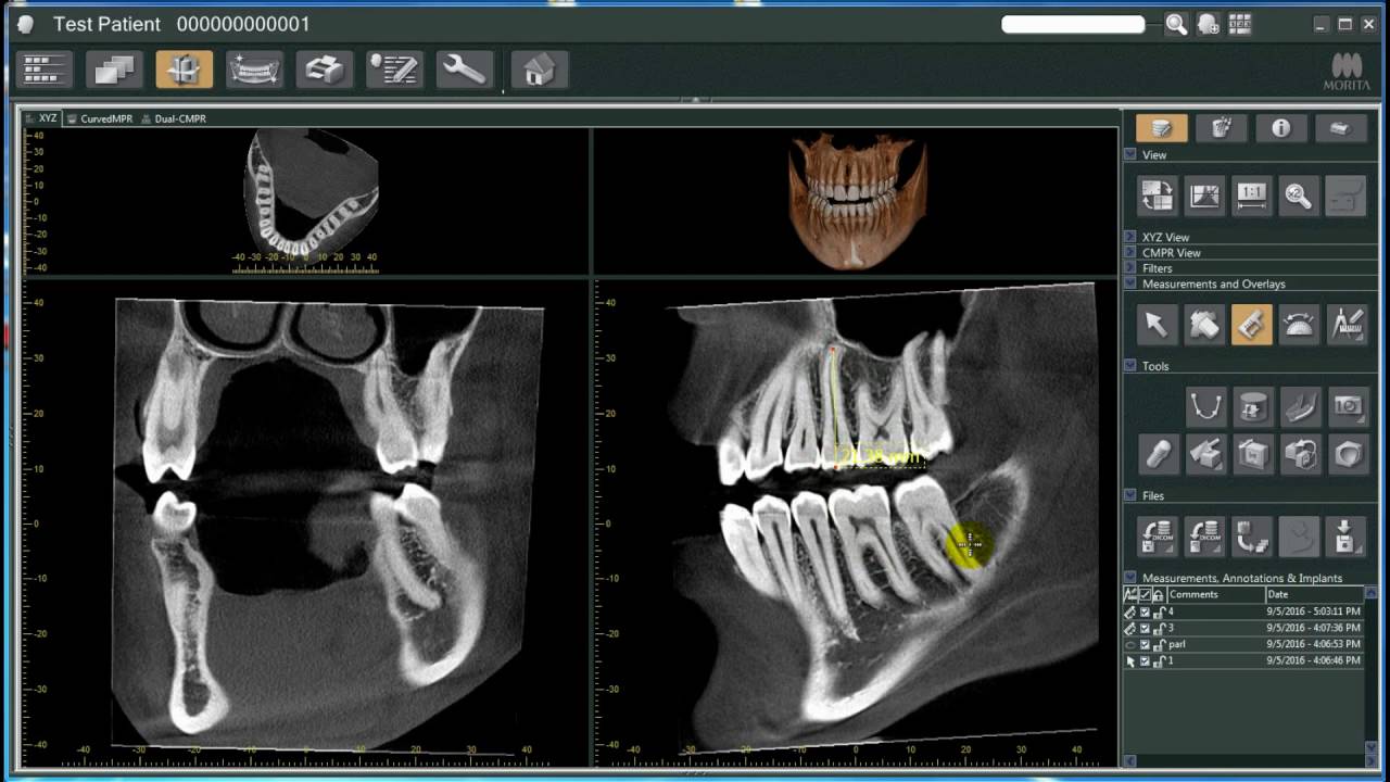
Orthodontists and dentists use Cone Beam Computerized Tomography CBCT which evolved from CAT. The improvements in imaging technology have helped in obtaining a near perfect image for accurate diagnosis. A 3D scan can help gain a better view of bone structures such as adjacent root positions in order to locate canals and root fractures as well as provide the ability to more.
3D dental imaging is rapidly becoming the standard of care in all of dentistry not just specialty fields. Imaging plays an important role in endodontics and is routinely utilized for the following diagnosis. The cone beam computed tomography is used to view the area of the head and neck in three dimensions and it is used to find the exact placement of implants the buccallingual position of impacted teeth to be removed and determination of the exact location of the mandibular nerve before surgery is done.
Dental x-rays help your dentist identify diseases and problems. The history of dental imaging began in the late 1800s with the development of the x-ray image. Digital radiography is a type of X-ray imaging that uses digital X-ray sensors to replace traditional photographic X-ray film producing enhanced computer images of teeth gums and other oral structures and.
Most states have laws that require inspections of dental x-ray equipment on a regular basis such as every _____years. Uses of digital imaging-To detect lesions diseases and conditions of teeth and surrounding structures-Confirm or classify suspected disease-Provide information during dental procedures root canal therapy instrumentation and surgical placement of. There are many types - or modalities - of medical imaging.
Many dental diseases and conditions have no clinical signs or sympstoms and are typically discovered. Radiographic features of 2D 3D and 4D imaging will be discussed. Hauser uses 3D scans to diagnose a wide variety of dental problems.
An offshoot of dental X-rays is 3D imaging. In the past two decades it has benefited from major advances in technology and has become an indispensable imaging modality due to its flexibility and non-invasive character. Dental x-rays can also help your dentist evaluate injuries to your face and mouth.
A variety of cases will be profiled to illustrate the impact of CBCT on diagnosis of resorption fractures periapical lesions and tooth anatomy. The use of dental x-ray equpiment is regulated by. Advantages and disadvantages of various imaging techniques in endodontic are presented.
Assessment of root morphology this may be important prior to dental extractions particularly for wisdom teeth. In the case of 3D dental imaging the advantages are clear granting practitioners and patients alike a better clinical experience. Dental professionals today are increasingly using digital dental radiographs digital X-rays to better detect diagnose treat and monitor oral conditions and diseases.
Not only is time saved in the development process reducing the amount of radiation by as much as 80. Barriers film speeds position of the operator and film processing. 3D Imaging Using CBCT.
Many dental diseases and conditions have no clinical signs or symptoms and are typically discovered only through the use of dental imaging. Describe the discovery of x-radiation. If you are a doctor who looks at the precision of this equipment and thinks its overkill for the patients you see it may be time to rethink that assumption.
Dental radiographs help dentists assess the development of teeth and bones in children. This course will describe the use of small field of view FOV CBCT in endodontic diagnosis and management of cases. A CBCT machine on the other hand uses a cone-shaped beam and a reciprocating solidstate flat panel detector which rotates once around the patient Figure 2 180-360 degrees covering the defined anatomical volume complete dental maxillofacial volume or limited regional area of interest rather than slice-by-slice imaging found in.
Conditions that are not visible in the oral cavity and to identify many conditions that might otherwise remain undetected. Radiographs enable the dentist to see. Periapical and cephalometric radiographs have.
Ultrasound is an inexpensive and widely used imaging modality for the diagnosis and staging of a number of diseases. Radiation health codes may include regulations pertaining to. Therefore imaging of these structures is one of useful diagnostic tools for clinicians to make decision treatment modality.
Medical imaging has led to improvements in the diagnosis and treatment of numerous medical conditions in children and adults. The uses of dental imaging are checking patients oral health and making it clearer to diagnose certain teeth problems such as cavities. Dental imaging enables the dentist to see conditions that are not visible in the oral cavity and to identify many conditions that might otherwise remain undetected.
Describe the purpose and uses of cone beam computed tomography.

Dental Radiology An Overview Sciencedirect Topics

Pdf Advances In Radiographic Techniques Used In Dentistry

Digital Versus Conventional Radiography In The Dental Office

Coconut Oil For Dental Health And Neurodegenerative Disorders Dental Health Dental Coconut Oil

Types Of Dental Radiographs And Their Uses Dentalnotebook

History Benefits Of Dental X Ray Imaging Artistic Touch Dentistry

Pin On Dental Favorites
What Is 3d Dental Imaging North Brand Dental Glendale Ca 818 244 7215

Orthopantomography Radiology Reference Article Radiopaedia Org


Comments
Post a Comment